√100以上 epimysium perimysium endomysium histology 115161-Epimysium perimysium endomysium histology
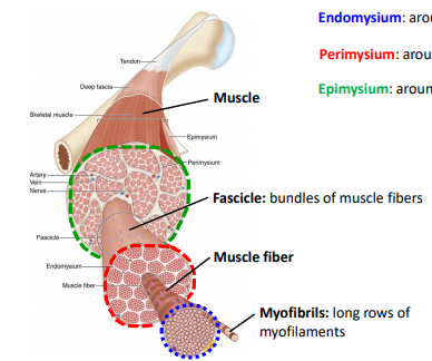
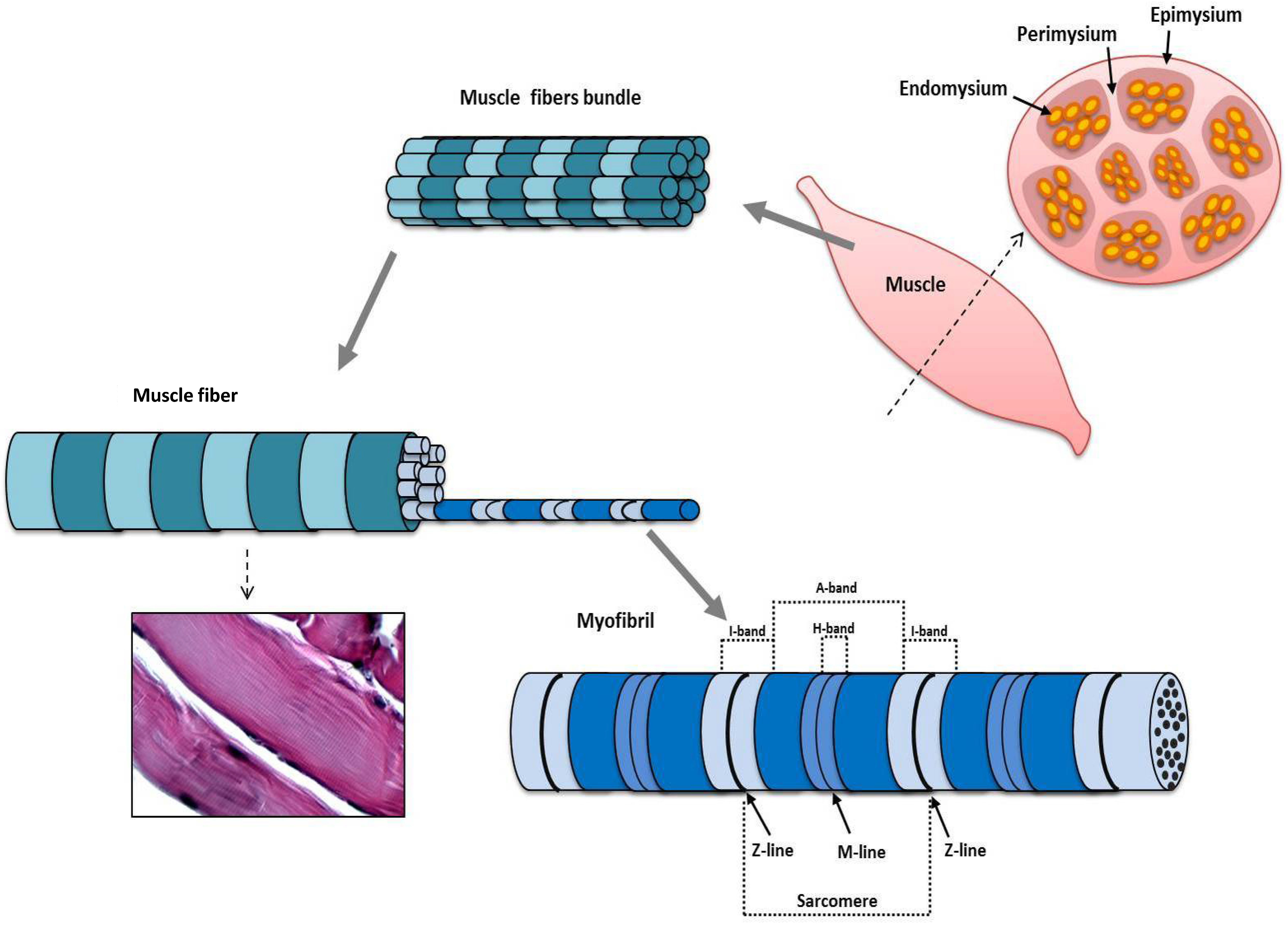
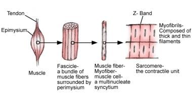
(2) separate the muscle into compartments; The most superficial layer of a muscle is the epimysium which wraps together numerous fascicles wrapped with perimysium Inside each fascicle are the individual muscle fibers wrapped around endomysium Within the muscle fibers is where the functional unit of the fiber exists the sarcomereHowever, the thickness and constitutent connective tissue fiber types of these sheaths varied regionally Near the articular capsule and disc, the endomysium, perimysium, and

Histology Of Muscle
Epimysium perimysium endomysium histology
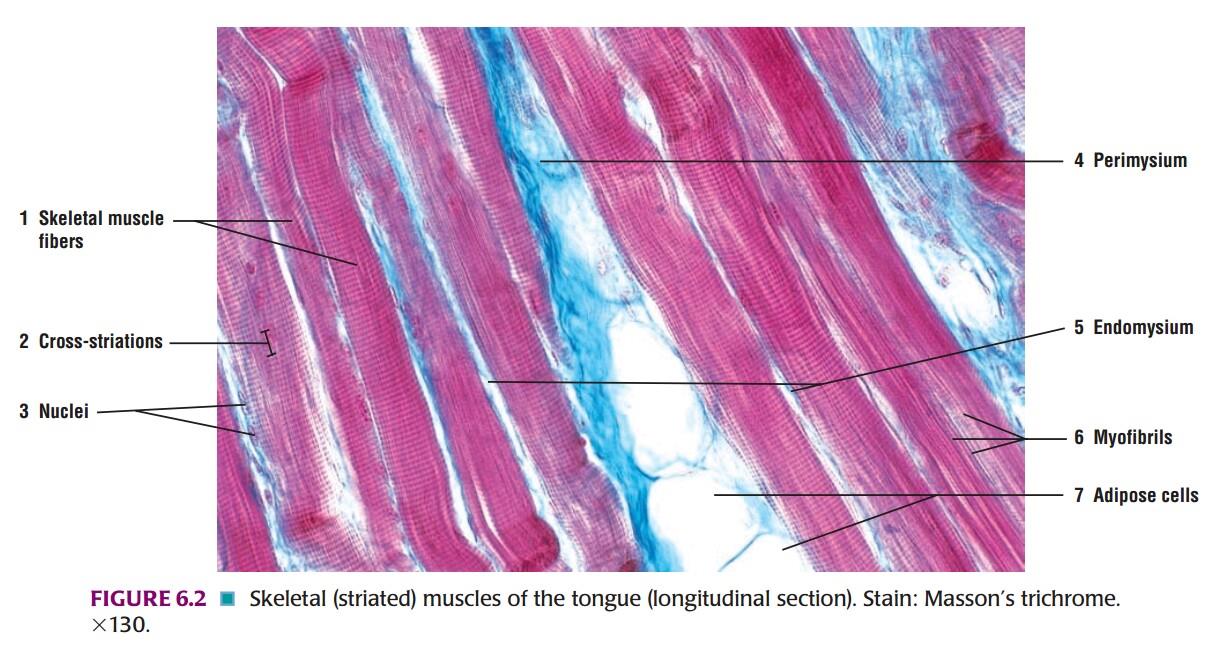
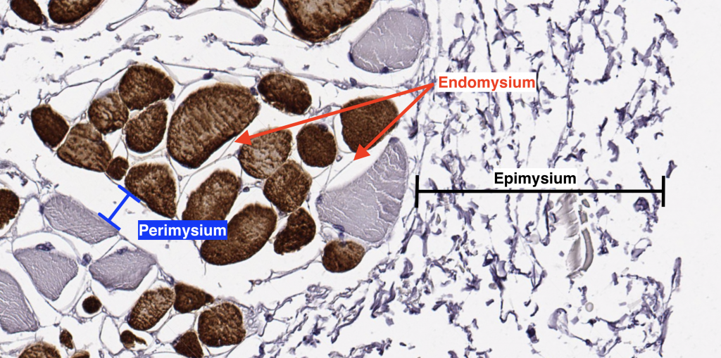
Epimysium perimysium endomysium histology-Perimysium encloses groups of fibers, defining fasciclesThis is a section of the tongue Begin by identifying groups of fasciculi cut in transverse section Where are the nuclei located within a cell?


Muscles Bioactive Collagen Peptides
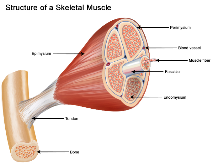
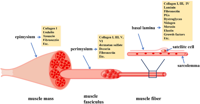
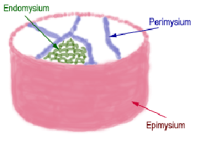
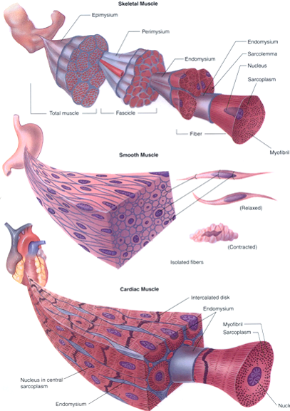
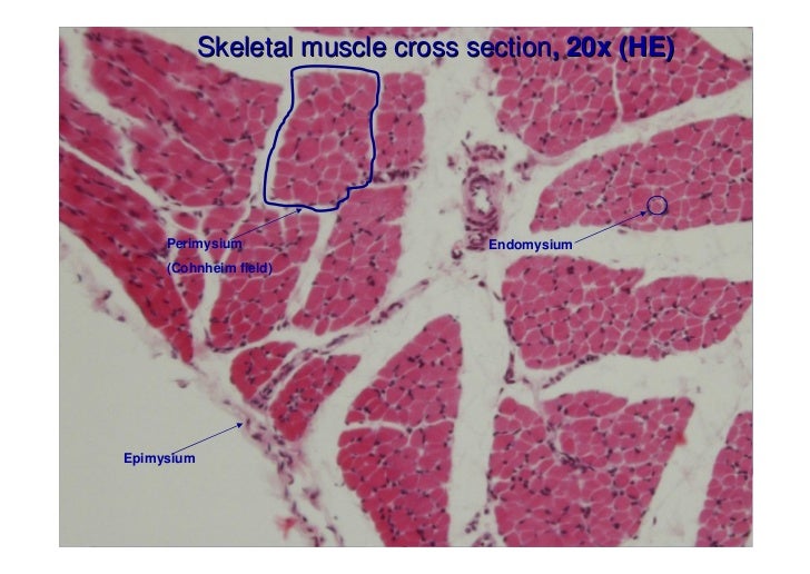
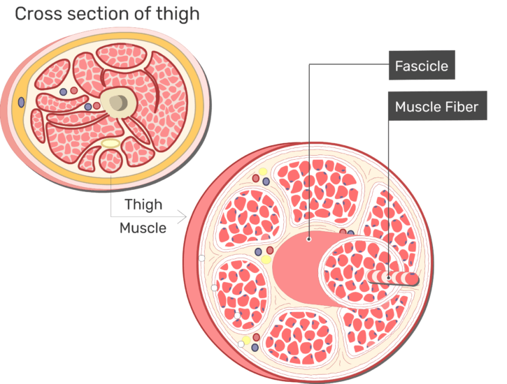
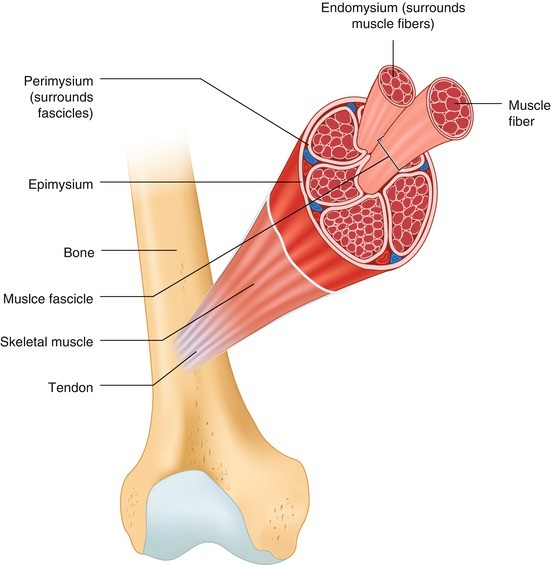
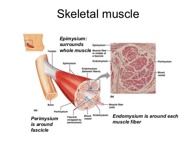
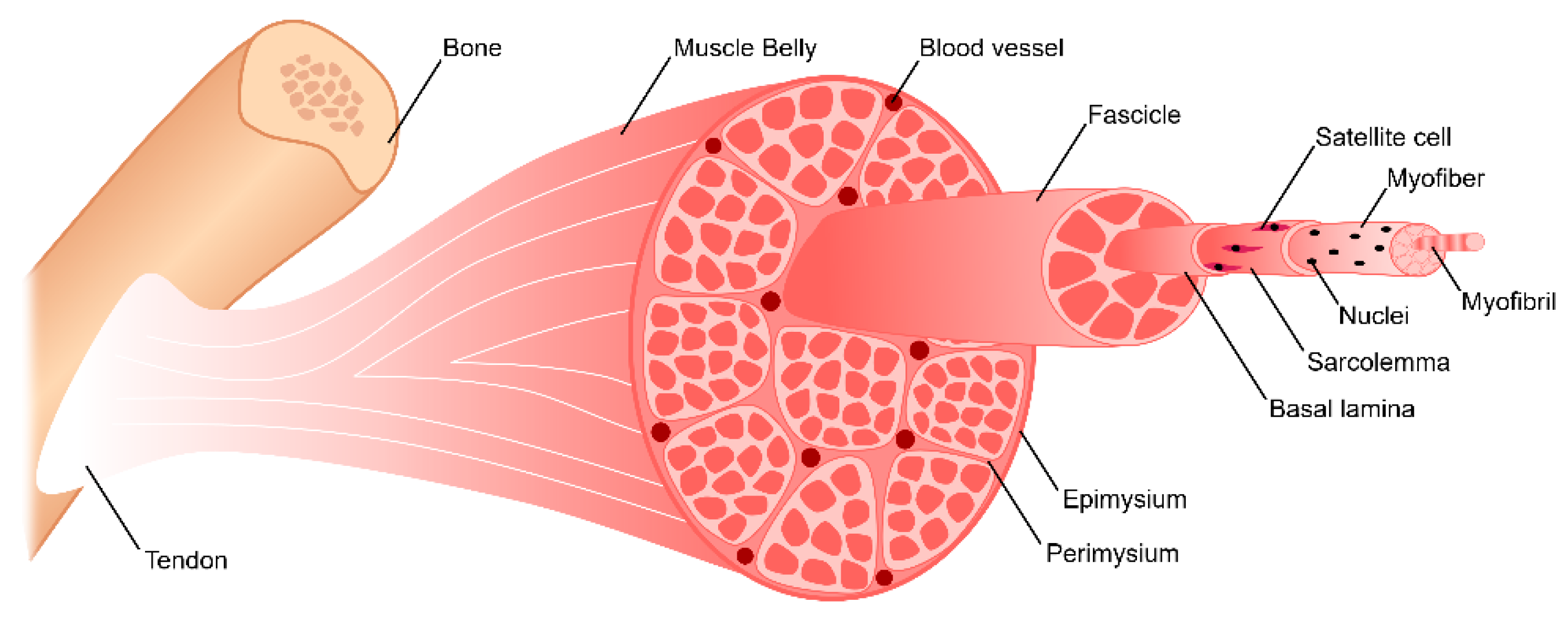
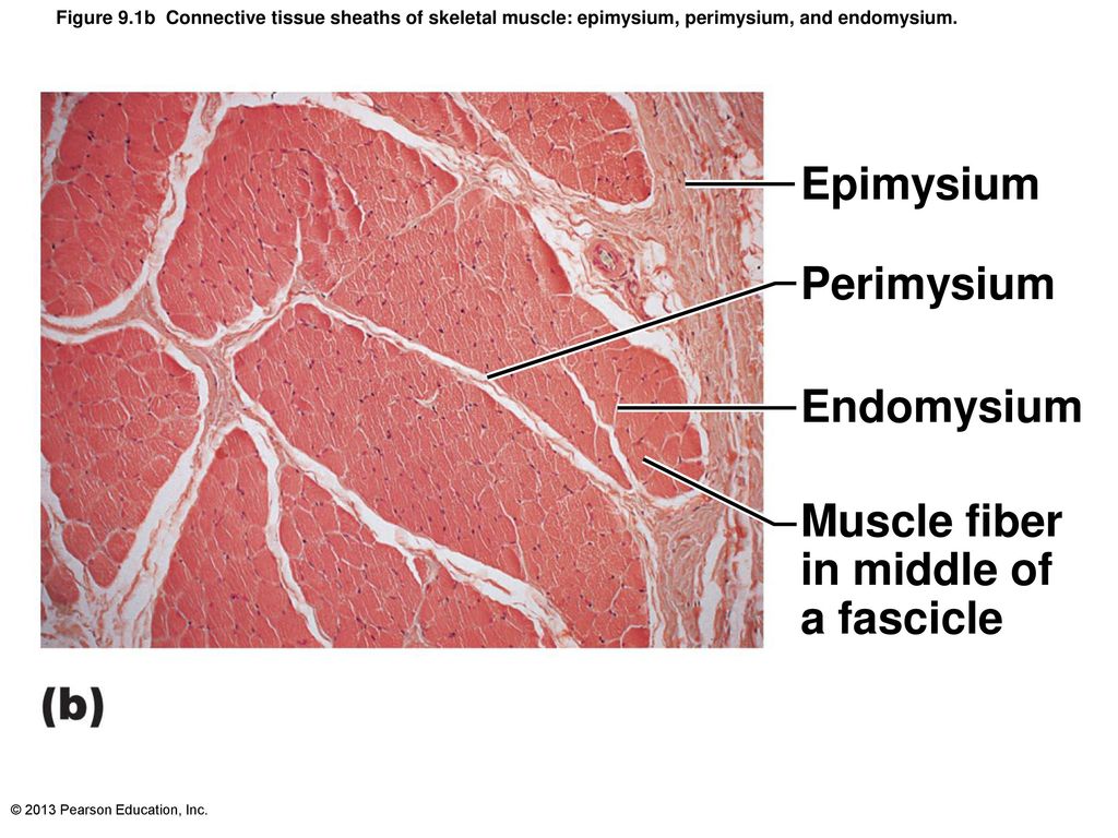
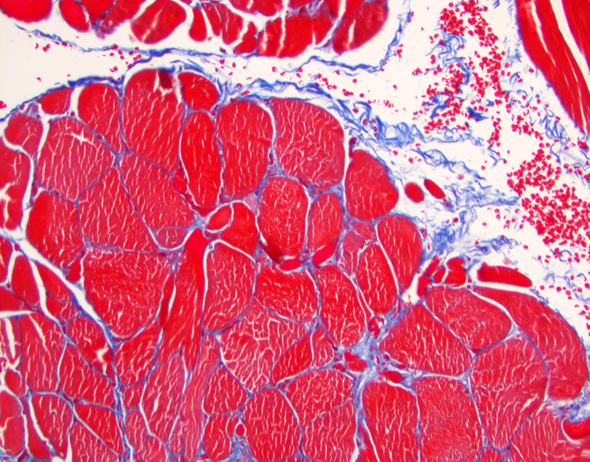
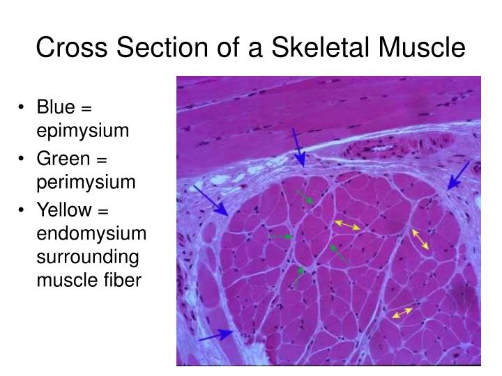
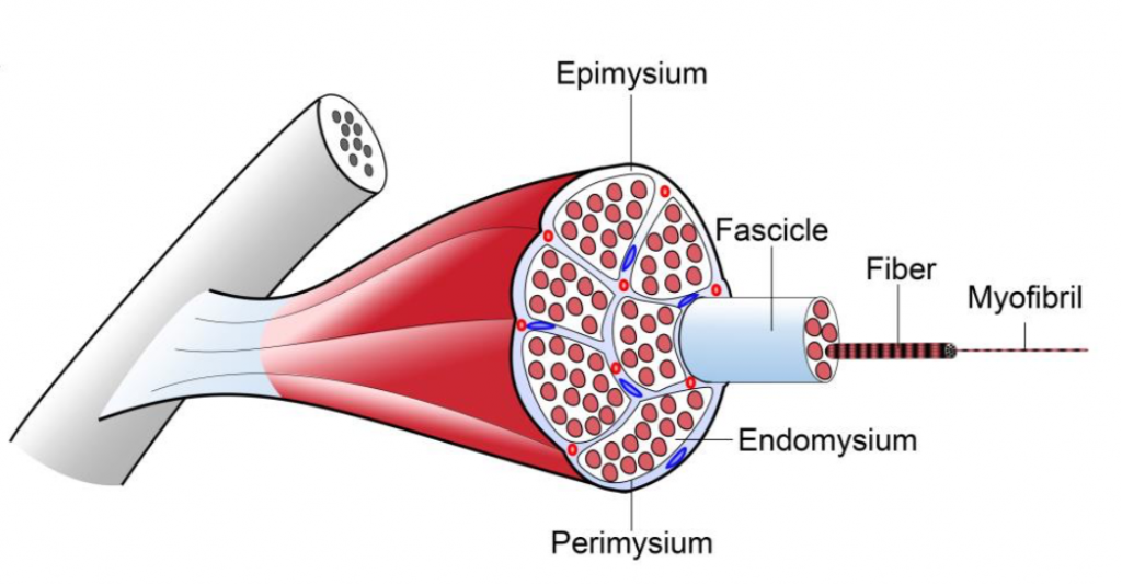
The whole muscle is enclosed by a dense connective tissue sheath called the epimysium (Epi greek for upon, mys greek for muscle) The muscle fibres are divided up into bundles of fibres called 'fascicles' Fibrous sheaths that surround the fascicles are called the perimysium (peri is greek for around) Blood vessels, the lymphatics and the nerves are all found in the perimysiumEpimysium is the specialized fascia located at the muscle Epimysium is a layer of connective tissue, which ensheaths the entire muscle It is composed of dense irregular connective tissue It is continuous with fascia and other connective tissue wrappings of muscle including the endomysium, and perimysium 1From the epimysium, thin collagenous septa extend inward to divide the muscle into a number of bundles or fasciclesThese septa are called the perimysium The perimysium is continuous with the endomysium that is a delicate connective tissue layer surrounds each individual myofibers
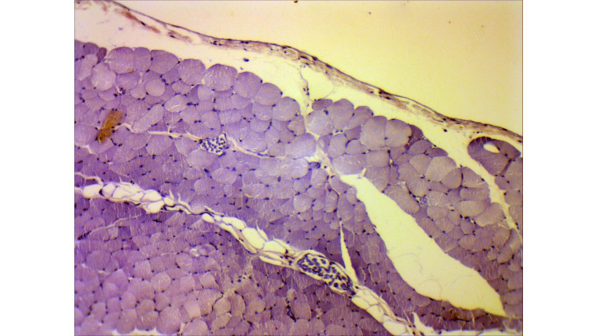
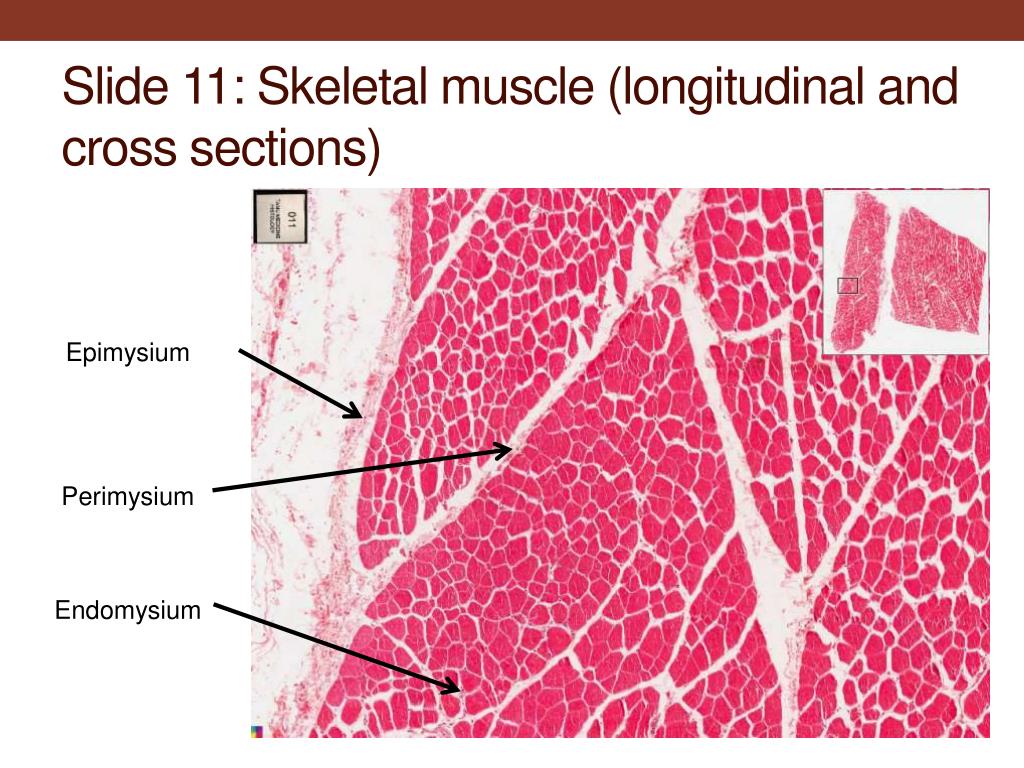
Groups of these fibrils form a muscle fiber, which is surrounded by endomysium Muscle fiber and muscle cell are synonymous Groups of individual cells that are surrounded by perimysium are known as fascicles and groups of these fascicles, surrounded by epimysium make up a muscle B Perimuscular Connective Tissue Here you will easily identify the epimysium, perimysium and endomysium of skeletal muscle fibers You will also find the ovoid nuclei in the skeletal muscle fascicles at the periphery Muscle slide images and videos I think these labeled images and diagrams of skeletal muscle histology were helpful to understand the basics of this muscle structure 1) Each muscle fibres is surrounded by delicate connective tissue that is called the endomysium 2) Individual fasciculi are enclosed by a stronger sheath of connective tissue called the perimysium 3) The entire muscle is surrounded by connective tissue called the epimysium This is illustrated by the schematic diagram below
Where can capillaries be found?The epimysium, perimysium and endomysium are noncontractile elements of a contractile tissue (muscle) Tendons are noncontractile, so they can be regular, because the tissue does not need to alter its length (in fact, for efficient function, a tendon should be as nonstretchy as possible)About Press Copyright Contact us Creators Advertise Developers Terms Privacy Policy & Safety How works Test new features Press Copyright Contact us Creators


Reading Php Lab


Animal Tissues Muscle Atlas Of Plant And Animal Histology
Epimysium A skeletal muscle is invested by three connective tissue layers that (1) bind the muscle together;The endomysium and perimysium were relatively thin and consisted mainly of reticular fibers The epimysium was thicker than the intramuscular sheaths and consisted of both collagen and reticular fibers;Anatomical terminology Perimysium is a sheath of connective tissue that groups muscle fibers into bundles (anywhere between 10 and 100 or more) or fascicles Studies of muscle physiology suggest that the perimysium plays a role in transmitting lateral contractile movements


How Muscle Structure And Composition Influence Meat And Flesh Quality


Seer Training Structure Of Skeletal Muscle
Can you identify the endomysium and the perimysium?Identify epimysium and perimysium With the 40X objective, examine muscle fibers cut in transverse and in longitudinal section and identify endomysium and blood vessels of various sizes (Figs 103 and 104) Where are the nuclei of these muscle fibers located?And (3) transmit the force of contraction to the muscle insertion Endomysium surrounds individual muscle cells;


Skeletal Muscle A 19 Diaphragm A 56 Muscle Skeletal Hmn Adult Ls A 57 Muscle Skeletal Fetal Ls Striation Large Elongate Cell Many Peripheral Nucleus Endomysium Ppt Download


Muscles Bioactive Collagen Peptides
How does the location of capillaries inThe mean strain for repairs with epimysium (104%) was significantly higher (p < 0001) than that for repairs without epimysium (73%) The mechanisms of failure were also different Among epimysium repairs, 15 stitches avulsed muscle transversely, and 10 stitches tore out longitudinally from the muscle In the nonepimysium group, 1 sutureE perimysium collagenous connective tissue that separates smaller bundles of muscle cells called fascicles F fascicle bundle of muscle cells bounded by perimysium G endomysium thinner layer of connective tissue that separates individual muscle cells III Skeletal muscle


Muscle Histology Ssn October 17 05 Presented By Tabassum Sardharwala Shannon Watkins Ppt Download


Muscle Tissue This Resource Is Licensed Under The Creative Commons Attribution Non Commercial No Derivative Works License Ppt Download
The interstitial connective tissue of muscle is subdivided into the epimysium (surrounds the entire muscle), perimysium (surrounds large angular fascicles divided into primary fascicles of 10–100 fibers), and endomysium (surrounds individual muscle fibers) The endomysium contains capillaries, nerve fibers, fibroblasts, and collagen fibrilsNear the articular capsule and disc, the endomysium, perimysium, and epimysium were all thicker than in other regions of the muscle and consisted of collagen, reticular, and elastic fibers


The Muscular Force Transmission System Role Of The Intramuscular Connective Tissue Journal Of Bodywork And Movement Therapies


Perimysium Histology Kcpc Org


Introduction To Mussels Human Anatomy Physiology I Dr
:background_color(FFFFFF):format(jpeg)/images/library/13584/skeletal-muscle-tissue_english.jpg)


Skeletal Muscle Tissue Histology Kenhub


Extracellular Matrix An Important Regulator Of Cell Functions And Skeletal Muscle Development Cell Bioscience Full Text


Home Php Lab


Muscle The Histology Guide


General Histology 2 Emedicodiary


Skeletal Muscle Consists Of Muscle Fibers Bound By Connective Tissue Download Scientific Diagram


X11 Histology Of Muscle Tissue Flashcards Chegg Com


Reading Php Lab


Muscle Fascia And Force Transmission Journal Of Bodywork And Movement Therapies


Muscle Tissue


Epimysium An Overview Sciencedirect Topics


Skeletal Muscle Histology Draw It To Know It


Skeletal Muscle Fiber Location And Arrangement


Histology Slides 1


1


Musculoskeletal System Springerlink


Diversity Of Extracellular Matrix Morphology In Vertebrate Skeletal Muscle Sleboda Journal Of Morphology Wiley Online Library
:watermark(/images/logo_url.png,-10,-10,0):format(jpeg)/images/anatomy_term/perimysium/ECXt7gUxRQFYokz6VI7Cw_Perimysium.png)


Perimysium Histology Modernalternativemama Com


Solved Label A Fascicle Perimysium Epimysium Chegg Com


Muscle


What Are The Epimysium Perimysium And The Endomysium Quora


Muscular System By Dr Shivarama Bhat Objectives To


1


Jaypeedigital Ebook Reader


Animalscience Tamu Edu


I Heart Histo F Is For Fascicle A Snuggle Of Cells Wrapped Up In A Connective Tissue Coat In Peripheral Nerves Fascicles Of Axons Are Surrounded By A Peri Neurium Coat


Perimysium Definition Anatomy


Mitochondria Again But This Time In Muscle The Ms Blog


Skeletal Muscle Physiology


Medicine Hack Endomysium Perimysium And Epimysium Definition Histology


Muscle Histology By Dr Armaan Singh


Muscle


Histology Practical Musculoskeletal Block Things You Need To


Histology Of Muscle


Histology 4000 Muscle Lecture Notes 6


Bioengineering Free Full Text Skeletal Muscle Tissue Engineering Biomaterials Based Strategies For The Treatment Of Volumetric Muscle Loss Html


Ppt Muscle Tissue Powerpoint Presentation Free Download Id


Cartilage Bone Muscle Histology Notes Medical Histology Jacobs School Of Medicine


Epimysium An Overview Sciencedirect Topics


Solved 1 1 N Oa 3 1 2 3 4 5 6 7 8 Blood Vessel Chegg Com


Onlinelibrary Wiley Com


What Is The Difference Between Epimysium And Fascia Quora


Histology Of Muscle


Fascicle


Histology Exam 2 Other Lectures


Anatomy And Cell Biology 3309 Lecture 3 Lab Notes Quiz 3 Oneclass


Chapter Opener 9 C 13 Pearson Education Inc Ppt Download


Endomysium Wikipedia


Skeletal Muscles Histology Fascicle Whole Muscle Muscle Fiber


4 Myofibril Stock Photos Pictures Royalty Free Images Istock


Muscle Tissue Sections


Histology Skeletal Muscle Epimysium Fascicle Of Muscle Cells Skeletal Muscle Medical School Muscle


Warm Up 1 Based On What You Know About Latin Root Words What Do You Think These Terms Refer To Sarcomere Sarcoplasm Myofibril Epimysium Perimysium Ppt Download


Jfmk Free Full Text Morphological And Functional Aspects Of Human Skeletal Muscle Html


Schematic Overview Of The 3 Components Of The Cardiac Interstitial Download Scientific Diagram


Chapter 9 Figure 9 1 Connective Tissue Sheaths Of Skeletal Muscle Epimysium Perimysium And Endomysium Diagram Quizlet


Histology Website Resource Ha9


What Is Muscular Tissue Histology Anjani Mishra


Human Muscle Epimysium Perimysium And Endomysium Muscular System Skeletal Muscle Muscle


Epimysium An Overview Sciencedirect Topics


Medicine Hack Endomysium Perimysium And Epimysium Definition Histology


1


Epimysium An Overview Sciencedirect Topics


Perimysium Histology Modernalternativemama Com


Pathology Outlines Histology Skeletal Muscle


Histology Of Muscle


Muscle
:watermark(/images/watermark_5000_10percent.png,0,0,0):watermark(/images/logo_url.png,-10,-10,0):format(jpeg)/images/overview_image/1186/YdTKKcsV7yJfzybtC3TNXA_histology-motor-unit_English.png)


Skeletal Muscle Tissue Histology Kenhub


Jaypeedigital Ebook Reader


Medicine Hack Endomysium Perimysium And Epimysium Definition Histology


Ppt Muscle Histology Powerpoint Presentation Free Download Id


Cartilage Bone Muscle Histology Notes Medical Histology Jacobs School Of Medicine


Medicine Hack Endomysium Perimysium And Epimysium Definition Histology


Muscle Tissue Petr Vanhara Phd Dept Histology Embryology Ppt Download


Skeletal Muscle Structure And Histology Overview Myofiber Types Histology Of Normal Skeletal Muscle


Medicine Hack Endomysium Perimysium And Epimysium Definition Histology


Vetmed Tamu Edu
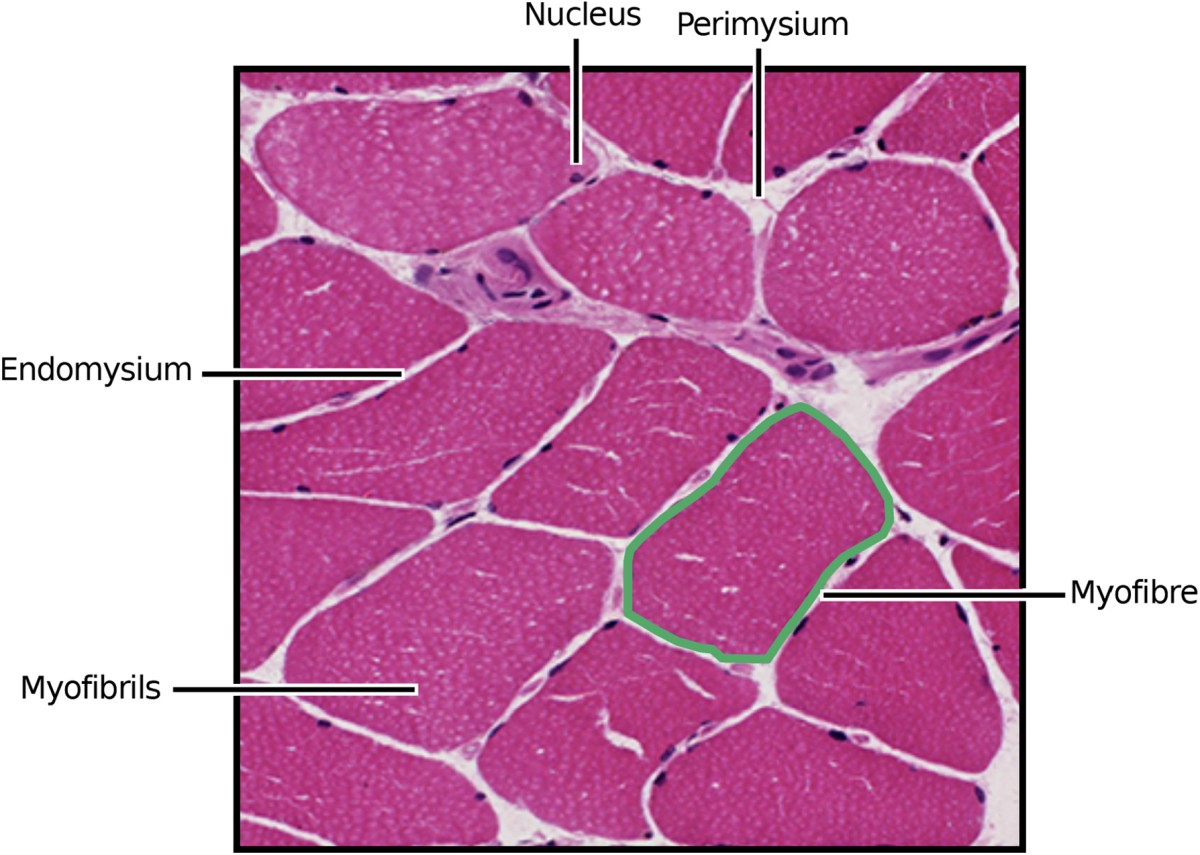


Myofibre Segmentation In H E Stained Adult Skeletal Muscle Images Using Coherence Enhancing Diffusion Filtering Bmc Medical Imaging Full Text


Muscle Tissue Sections


Normal Histology


Muscle Skeletal Muscle Transverse Cut A B C D A Epimysium B Perimysium C Endomysium D Fascicle Ppt Download


Sokvetjournal Net


Histology Of Muscle


Skeletal Muscle Histology Slide Identification And Labeled Diagram Anatomylearner The Place To Learn Veterinary Anatomy Online


Histology Muscle Flashcards Quizlet


Epimysium An Overview Sciencedirect Topics
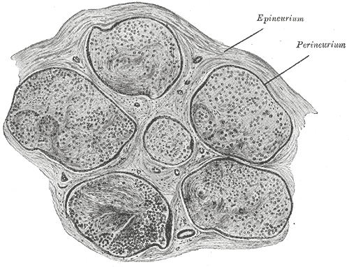


Perineurium Wikipedia


Muscle Histology Youtube


Schematic Diagram Showing Arrangement Of Extracellular Matrix Ecm In Download Scientific Diagram


Usmle Anatomy Chapter 4 Muscle Tissue Flashcards Quizlet
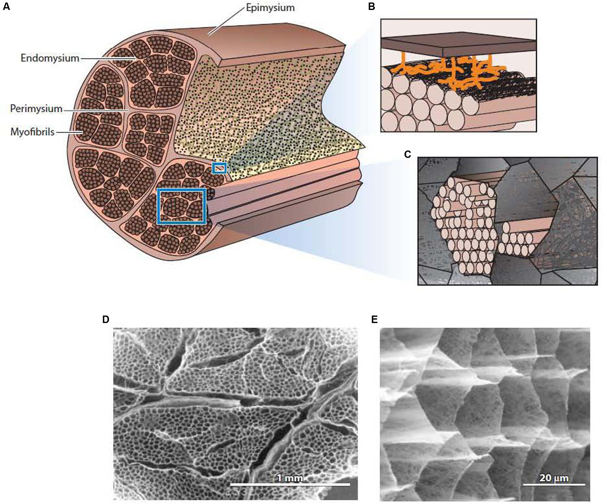


Frontiers The Structure And Role Of Intramuscular Connective Tissue In Muscle Function Physiology
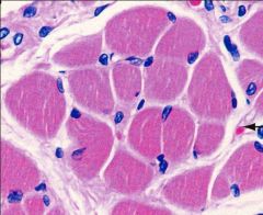


Histology Exam I Part 2 Flashcards Cram Com


Muscle Histology Prelab
コメント
コメントを投稿